
Discover a Wide Range of Health Care Services
We provide compassionate, dedicated specialty medical care at every one of our locations.

We're how. You're why.
Finding the right physician is key to establishing a medical home so that care is received in the right place, at the right time, in the manner that best suits your needs. Patient ratings are just one of the many ways we uphold our commitment to providing excellent, patient-centered care to San Antonio and South Texas.

From Our Patients
-
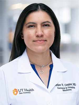
Dr. Clapper takes her time with me during the visit. I don't feel rushed and she listens to me attentively. She exhibits a genuine interest in my medical care. She is a great role model for other physicians.
UT Health San Antonio Patient
about Amber Clapper, MD -
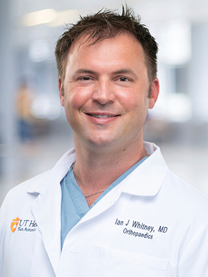
Dr. Whitney was outstanding -- professional, had reviewed my case and MRI, had a plan in mind but discussed it fully with me as a team; I would rate much higher than a 5 if I could.
UT Health San Antonio Patient
about Ian Whitney, MD -

As soon as I left the office I was raving about my new PCP to all of my friends. She made me feel heard and was patient with me as I explained my concerns to her. She was so kind and made me feel taken care of. At office visits in my past I would have to fight to have the doctor take me seriously. Dr. Perez took my concerns seriously and listened to me. We discussed all of my concerns and made a plan for my healthcare. This was the best doctor's visit I've ever had. It was easy to take speak with my doctor and I felt we made a good connection. I look forward to being a patient here for a long time.
UT Health San Antonio Patient
about Mayra Perez, MD -

Dr. Chan listened and elicited more info with appropriate questions, performed a thorough physical exam and provided a rational, easily understood plan for further studies and interval treatment. Very reassuring.
UT Health San Antonio Patient
about Cassie Chan, MD -
I have never had a doctor and nurse to take the time to listen intently and provide information as much as the staff in this clinic have provided- I am so thankful to have found this clinic!
UT Health San Antonio Patient
about Jason Brownell, MD
We promise to be friendly and welcome you warmly, provide outstanding medical expertise, be compassionate every step of the way, give undivided attention to deliver a personal experience, be sensitive to your needs and respectful of your time... always. This is our promise and our passion.
UT Health Physicians offers the region's most comprehensive services. In every discipline, our team of highly trained and board-certified physicians and surgeons work together to provide the highest quality of compassionate, patient-centered care.


We're here to be your medical home - full of trusted, experienced primary care providers who focus on internal medicine, family medicine, geriatric care and pediatrics. Our team is formally recognized for the way we care for our patients and for being their direct connection to San Antonio's most experienced doctors in every medical specialty.
Our mission is to decrease the burden of cancer in San Antonio, South Texas and beyond. We bring South Texas a level of exceptional care that is comparable with the nation’s most respected programs. More patients put their trust in our program because we have a unique understanding of our community’s cancer care needs. We excel in delivering advanced therapies


MyChart: Access from anywhere
UT Health Physicians offers MyChart for secure access to your medical records from your computer or smartphone. MyChart users can schedule appointments and have direct access to their providers.